Removing a huge tumor as big as a “second head” for patient
20/06/2023 14:56
NDO – Despite of facing the risk of profuse bleeding causing the patient to die right on the operating table or post-op complications such as leaking cerebrospinal fluid or severe infection, the doctors of Viet Duc University Hospital have determined to perform a particularly difficult surgery. They initially successfully removed a giant neurofibromatosis tumor larger than the head of a female patient.

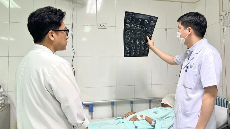
The doctor examines the patient after surgery.
Born in a family with all 3 siblings suffering from an incurable genetic disease causing tumors to grow all over their bodies, Ms. P. (40-year-old, Thai Binh) is the one who suffers the most with a huge tumor on her face.
Due to their difficult circumstances, there was no way to be accessed to the health care, so the tumor on her face kept growing more and more. The nose and upper lip were enlarged to more than 10 times the original size. Over half of her face has developed into a tumor as large as the rest of her head.
Her entire eyeball was pushed out of its socket and the blood vessels proliferated on the tumor like a fire dragon’s eye. Only until one day when the tumor grew too big, became swollen, painful, tense and she thought it was about to burst, the three siblings decided to bring their youngest sister to the medical facility for treatment.
Associate Professor, Dr. Nguyen Hong Ha, MD, PhD, Head of Maxillofacial-Plastic-Aesthetic Surgery department, Viet Duc University Hospital, said that through examination and diagnostic imaging, doctors diagnosed the patients with giant neurofibroma in the left head region.
“Congenital tumor, in addition to proliferating outward, also destroys a large part of the sphenoid bone of the skull and orbit. A large part of the tumor grows outward from the brain, causing the eyeball to fall out of the socket, the tumor communicates directly with the brain.
Not to mention, due to the characteristics of this giant tumor, the tumor is supplied with blood by a series of interlaced vascular systems inside and outside the skull, making the tumor always beat in rhythm with the heart and always hotter than other areas of the body up to 1 or 2 degrees Celsius making treatment very difficult,” said Dr. Ha.
According to Dr. Ha, the surgery for the patient may have a potential risk of profuse bleeding, causing the patient to die right in during the operation or the patient may experience meningitis or a leakage of cerebrospinal fluid after surgery.
The maxillofacial plastic surgeons conducted interdisciplinary consultations throughout the hospital with specialists in neurosurgery, diagnostic imaging, anesthesiology, resuscitation, blood banks… to formulate a detailed surgical plan and carefully discussed by experts.
Dr. Ha suggested that surgical treatment for this patient may have to be carried out in multiple stages to ensure maximum safety for the patient.
In the first surgery, the maxillofacial surgeons will have to make careful calculations to be able to resect almost 70-80% of the tumor out without losing too much blood. Next, neurosurgeons will have to remove the part of the tumor communicating with brain tissue, placing artificial metal sheets to reconstruct the base of the skull, separating the brain tissue from the tumor. Then, the doctors will carefully stop the bleeding and reconstruct with stitches to close the resulting lesion.
The surgery took more than 8 hours. To reduce the risk of massive blood loss, radiologists had to perform selective nodal intervention on the blood vessels supplying the tumor prior surgery.
However, because the internal carotid artery which also supplies blood to the tumor is not allowed to be intervened with as it is the main artery that supplies blood to the brain, during surgery, every time the skin was cut, the blood from the skin edges still gushed out like a stream, the doctors had to work very hard to stop bleeding with each centimeter.
To ensure the safety of the patient during operation, the doctors of the Anesthesia Resuscitation Center had to apply many state-of-the-art machinery systems from the extremely difficult stage of intubation to monitoring the resuscitation during and after surgery.

Doctor Dao Van Giang, MD, PhD, Deputy Head of Maxillofacial-Plastic-Aesthetic Surgery Department, Viet Duc University Hospital – a member of the surgical team said: After more than 2 weeks of intensive postoperative care at the Department of Maxillofacial, Plastic and Aesthetic Surgery, Ms. P. was able to eat and drink through her mouth, sit up on her own, and walk gently.
“Most of the tumor has been removed, she is relieved of a huge burden on her face and ready to return to her daily life. The wound will be taken care of with frequent dressing changes while we wait for the patient to make a full recover before proceeding with the next surgery in which we will operate on the remaining tumors of the lips and nose,” said Dr. Giang.
According to Associate Professor Nguyen Hong Ha, MD, PhD, neurofibromatosis is a disease caused by genetic mutations, particularly changes in the gene structure on the 17th chromosome. This change affects the gene that regulates nerve fibers leading to uncontrollable tumor growth in a benign or even malignant direction.
Tumors develop in many forms on the body. From patients with hundreds of tumors like warts all over their body, to giant tumors like bear arms, elephant legs, horse’s mane nape, giant tortoise shell back.
In addition to the case of largest neurofibromatosis tumor operated on (weighing 45kg) by Vietnamese doctors: patient Nguyen Van S. (Nghe An), this case was also one of the largest neurofibroma of the head, face and neck ever recorded in Vietnamese medical literatures.
Associate Professor Ha recommends that patients who misfortunately suffer from any congenital disease should go to meet the doctor as soon as possible, the surgery will be much simpler and less dangerous than when the patient show up late. If not treated early on, large tumors can burst at home causing death or some tumors can turn malignant which will be much more difficult to treat.


