A woman was pierced by a 12-centimeter long piece of wood penetrating through her eye and brain
16/04/2021 07:21
When going to the forest for harvesting bamboo, a 40-year-old woman, native of Bac Kan province was suddenly hit on her head by a tree branch.

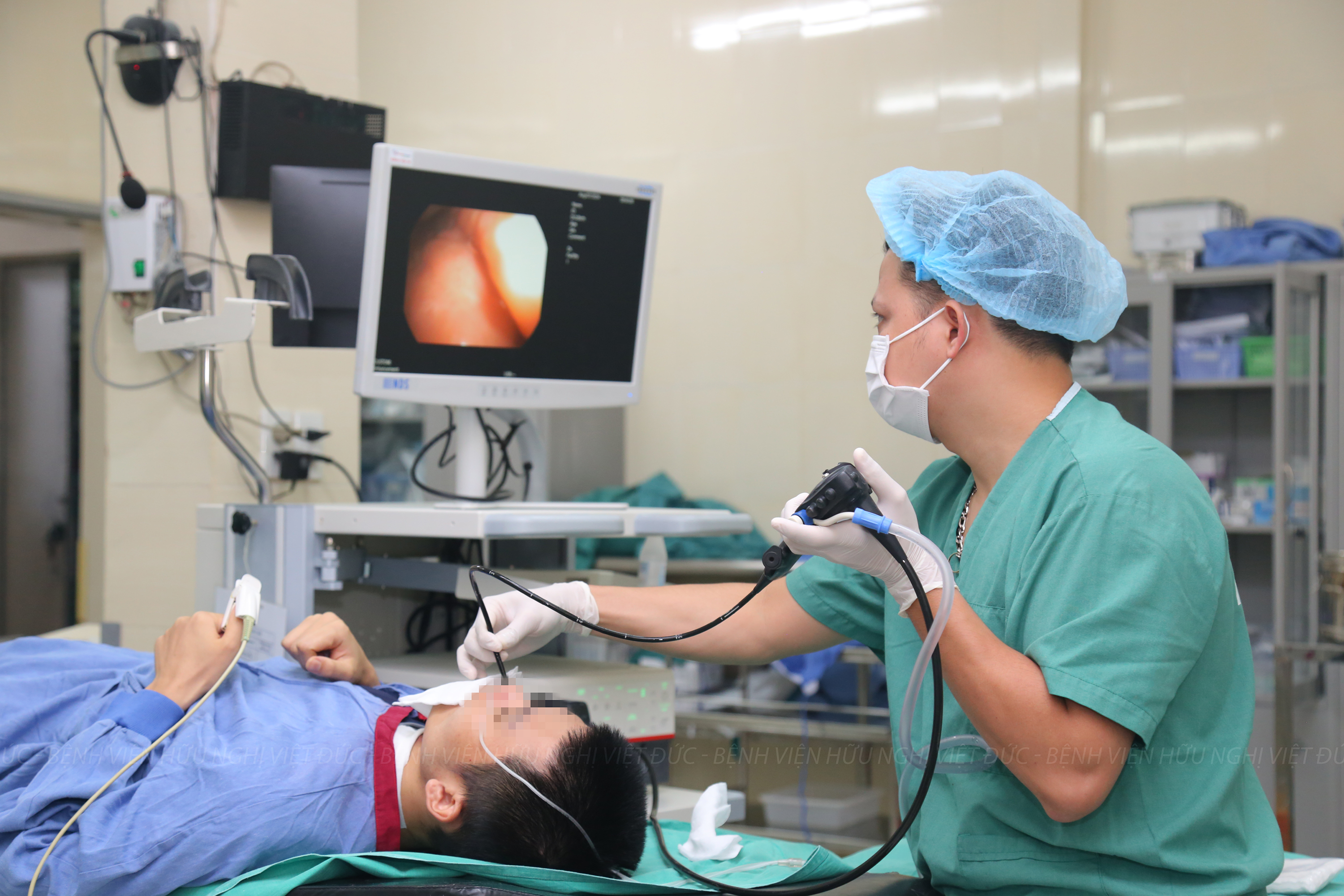
Doctors were performing an operation to remove the foreign body from patient
After the accident, the patient lost consciousness at scene, then her family quickly carried her down the mountain to a district hospital.
There, doctors provided the first aid but they did not good enough equipment so it’s not possible to determine whether there was a foreign body in patient’s brain or not. So, in the same day, the patient was transferred to Viet Duc University Hospital.
Dr. LE Hong Nhan, PhD., MD., Vice director of Neurosurgery Center, Head of Neurosurgery II Department shared: On admission, the patient was alert without neither paralysis nor fever; her right eye was bloody swollen with exposed intra-ocular tissues; the eyeball was collapsed suspecting an orbital fracture. Clinical examination and diagnostic imaging found a foreign body that was likely to be a long piece of wood penetrating into eye socket through cavernous sinus to posterior cranial fossa with signs of bleeding and air collection around.
A cerebral angiography and a MRI were performed to exclude cerebral vascular injury. Cranial images showed a foreign body penetrating into the right eye socket through optic foramen, exterior wall of the right cavernous sinus, superior border of petrous bone and cerebellar tentorium to the cerebellum just beside the brainstem causing hemorrhage and air collection along the passage of the foreign body.

Successful operation to remove a 12-centimeter long piece of wood penetrating into patient’s brain. Source: Image provided by VDUH
The patient was consulted under the direction of Professor TRAN Binh Giang, PhD., MD., Director with leading experts of hospital and Dr. BUI Thi Huong Giang from National Institute Of Ophthalmology decided to remove the eye ball together with the foreign object inside.
The operation was done on March 29th, 2021 to remove the eye ball and the foreign object inside.
To prevent and control potential injury of major vessels in the skull base, while removing the foreign object, neurosurgeon team did explore the right common, internal and external carotid arteries. Intravascular intervention team from Diagnostic Imaging Department, Viet Duc University Hospital also well prepared all equipment and materials to perform per-operative embolization.
Anesthesiologist team prepared all infusion lines and solutions for resuscitation in case of massive hemorrhage. This operation for removing the foreign object was performed under surgical microscope via orbital approaching after removing the eye ball.
The foreign object was a 12-centimeter long piece of wood with pointy end; its internal top inside the brain was about 0.5 cm wide; its remaining top stuck in the skull bone was 1 cm thick.
So far, patient continues to be monitored and cared to prevent the post-operative infection and hemorrhage.


