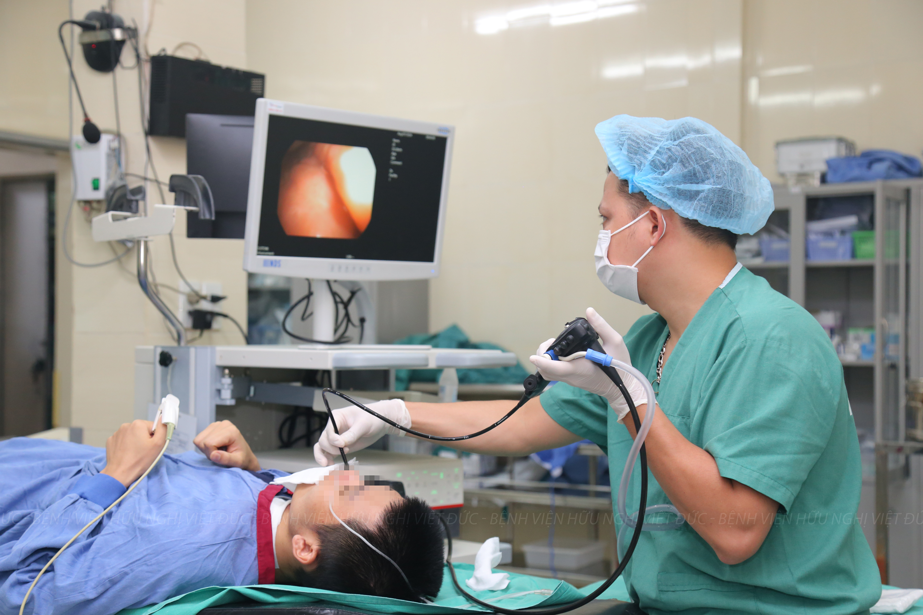
Early damage of cardiac, nerves, cancer … will be detected thanks to this equipment system
25/09/2019 10:14
With a series of devices for the most advanced paraclinical indications opened at Viet Duc University Hospital on September 12, more patients will be screened for early detection, timely treatment, and contribution in the quality of treatment for patients…
Accordingly, the newly equipment at Diagnostic Imaging Department of Viet Duc University Hospital includes 512 slices CT, 3.0 MRI and DSA scanners to respond to the needs of patients
Speaking at the ceremony, Prof. Tran Binh Giang – Director of Viet Duc University Hospital said, with a team of leading surgical center in Vietnam, this system will support the hospital to constantly assert its reputation in examination and treatment for the people.
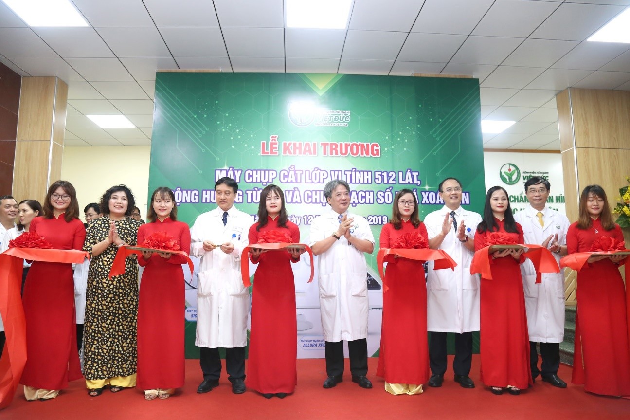
Viet Duc University Hospital launching the world’s leading equipment group in diagnosis and treatment
Accordingly, difficulties in detecting and treating cardiovascular, neurological, musculoskeletal, abdominal, breast, thoracic and cancerous lesions will be overcome. Clear images, high resolution, help doctors detect disease early and have appropriate solutions.
“In particular, not only fast capture time, this medical equipment system also limits X-rays for both patients and medical staff to more than 80%. This is a breakthrough in diagnosis and treatment of hospital. Hence, hospital requires physicians to make the most of modern equipment in treatment, as well as in training, scientific research and international cooperation to serve the quality of medical examination and treatment for people.” – Director Tran Binh Giang informs.
Prof. Tran Binh Giang also hopes , this state-of-the-art equipment system will both help reduce patient load and improve the quality of diagnosis and treatment for patients. Dr. Vu Hai Thanh, second specialized degree – Chief of Diagnostic Imaging Department, Viet Duc University Hospital shares, with this new equipment, when patients are assigned to perform one of the techniques of 512 slices CT system, 3.0 MRI and DSA machine will benefit. Due to the advantages of these devices such as: due to the fast capture time (only taking the whole heart in 1 heart rate, or the heart rate over 70 still taking good shots), it not only saves the patient time and time. manage the side effects when using contrast medicine, medicine to stabilize blood pressure for people with medical conditions of blood pressure but also help reduce radiation dose affecting the body.
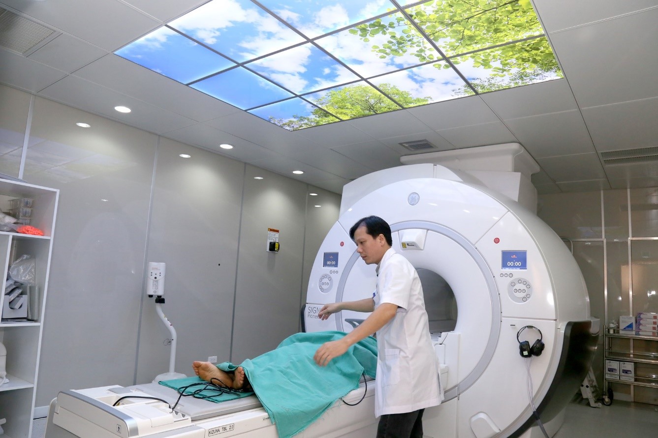
The introduction of modern equipment for diagnosis and treatment at Viet Duc University Hospital could contribute for improving the quality of medical examination and treatment
Experts on diagnostic imaging also said that, along with reducing the amount of radiation for patients, the machine produces sharp images for intensive intervention. In addition, there is a mode of 3D image reconstruction, angioplasty to measure the size, choose the approach method to damages. This device is suitable for many diseases, especially emergency liver injury, kidney rupture can be conserved treatment.
Previously, the traditional method was to perform surgery to stop the bleeding, or hepatectomy, now there are vascular interventions, just need to thread in the bloodstream and pump substances that stop the bleeding. Previously, the procedures lasted up to 4-5 hours, now with smart devices it only takes 1-2 hours to complete, including complex injuries.
About the DSA scanner will erase all other parts, only shows the blood vessels, so the doctor can easily diagnose and handle the damage. Images are zoomed in more detail when stenting cerebral vessels, coronary arteries; The device automatically measures the narrowness and length of the circuit so that the doctor can select the appropriate stent size and place it in the correct position.
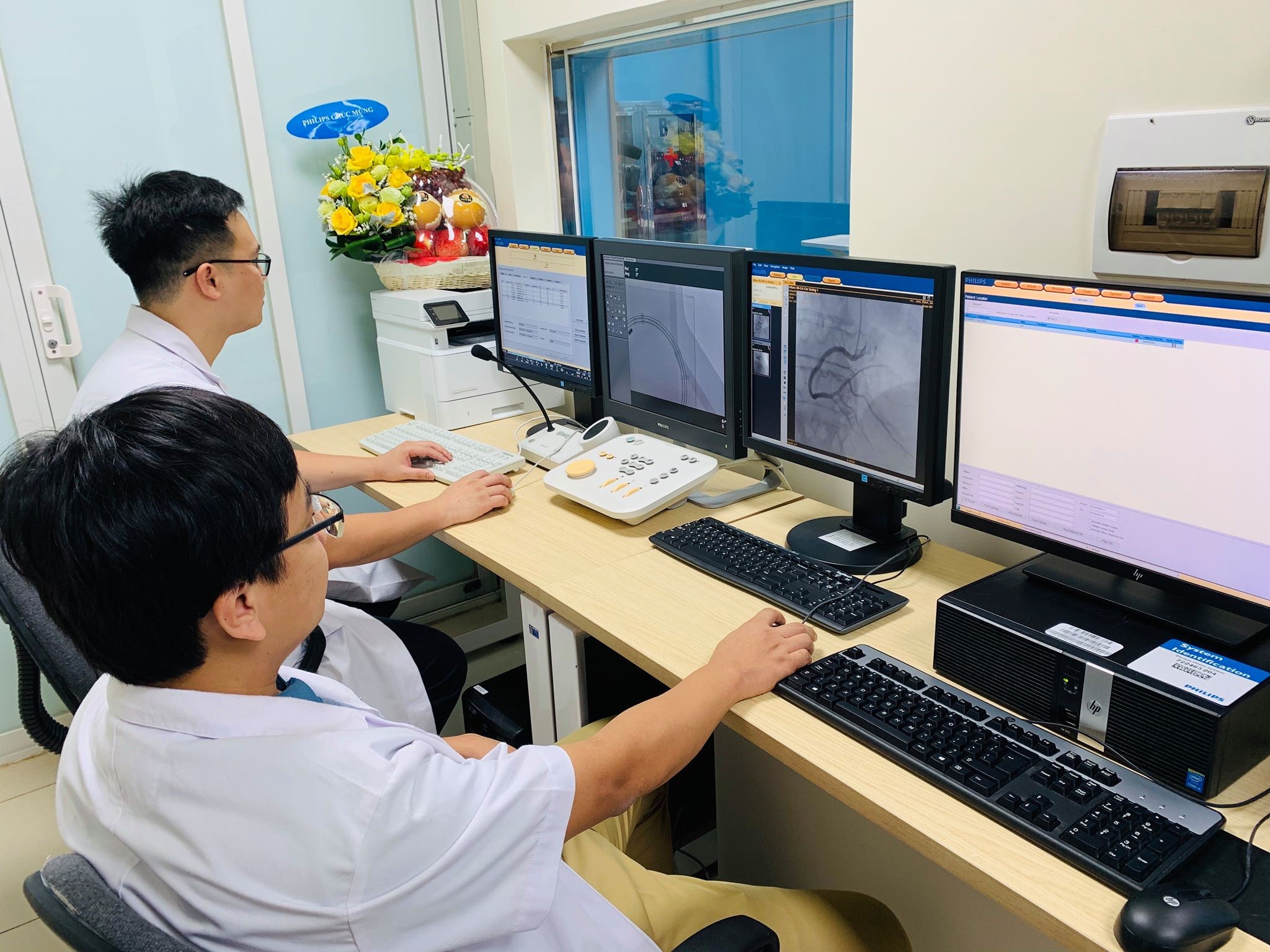
DSA scanners with many advantages have been retrofitted at Viet Duc University Hospital
According to Prof. Nguyen Huu Uoc, with cardiovascular diseases, 512 slices CT system, 3.0 MRI and DSA have made breakthroughs in diagnosis and treatment. The images taken are clearly displayed, fast shooting time, X-ray restrictions for both patients and medical staff.
“In addition, the diagnostic imaging system has replaced the method of exploration to expand the appointment and early diagnosis of diseases other than cardiovascular diseases” Prof. Nguyen Huu Uoc said.
With the diagnosis and treatment of diseases related to bone and joint injuries,, Ass.Prof. Nguyen Manh Khanh – Chief of Department of upper extremities surgery and Sport medicine, Viet Duc University Hospital said, Currently, MRI 1.5 helps to read damage to tendons, muscles, ligaments well, but with injuries of cartilage, condylar lesions, tibia tuberosity … is limited.
While the general trend is osteoarthritis, trauma, cartilage damage … is increasing, so early detection of small injuries to tendons, muscles, cartilage … is essential.
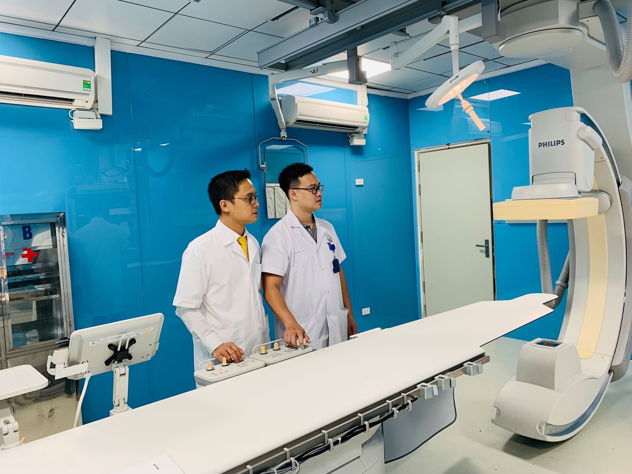
Modern equipment system is put into service for patients at Viet Duc University Hospital
Equipped with a 512 slices CT system, the 3.0 MRI will contribute to a dramatic change in diagnosis and treatment. And the retrofit of digital background scanners (Viet Duc University Hospital now has one) will increase the strength of doctors’ diagnosis and detection process.
Thái Bình Journalist / Health and Lifestyle


