Eliminating giant meningeal tumor for a patient with modern navigation system and Harmonic scalpel
11/08/2020 14:16
The 57-year-old woman has just been revived by doctors at the Viet Duc University Hospital with modern navigation technology to correctly locate the giant meningeal tumor of sphenoidal bone and a special help from the ultrasonic scalpel (Harmonic scalpel). This is also a warning not to look down on even benign tumors.
Doctor Vu Quang Hieu – Department of Neurosurgery II, Viet Duc University Hospital – is visiting patients after surgery.
Patient named D.T.D, 57 years old, living in Tu Ky, Hai Duong, was hospitalized with giant meningeal tumor located at sphenoidal bone, up to 62x59x50mm in size which has compressed and reduced Ms.D eyesight for a long time.
As the person directly operating the patient D, Doctor Vu Quang Hieu said: Meningeal tumor has attached the entire left sphenoidal bone of the patient’s brain. Although most meningeal tumors are benign, they are very proliferative. Blood vessels include two main sources: the artery in the cranial base of the external carotid system and the branches of the artery from the brain which related to the inner carotid artery system.

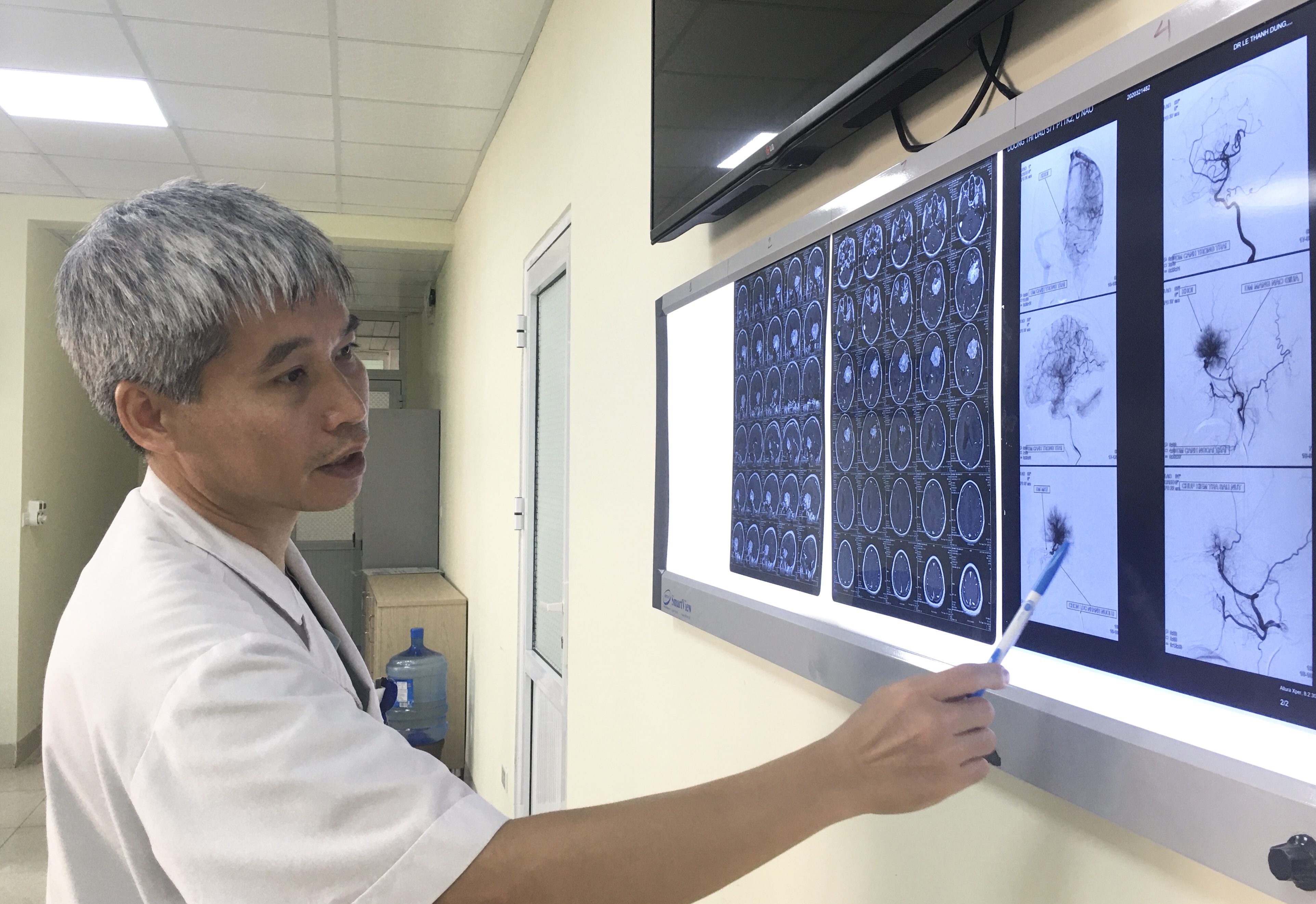
Doctor Vu Quang Hieu checked the x ray of patient
Because of the specific location of the tumor at sphenoidal bone, wrapping around the entire branch of the carotid artery including the intracranial carotid artery, which divides into the anterior cerebral artery, the medial artery, and the posterior catheter. The whole adjacent structures include: nerve number II, nerve number III, nerve and nerve near the eye. This tumor usually develops slowly, so when the patient comes to the hospital, it has grown very large.
Because of this structure, anatomy location along with tumor development, the whole tumor may not invade the vessel, but still attach to the cerebral vessels in the base of the skull from the carotid artery to the anterior cerebral artery, the middle cerebral artery, the posterior catheterization, nerve structures, II nerves, and III nerves which leaded to be difficult to remove by surgery. Especially, doctors wouldn’t find the nerve structures that need to be protected until they have dissected and taken out all the tumor inside.
To be able to remove all tumors, doctors must coordinate between many specialties. Doctors in Department of Diagnostic Imaging had captured arteries and embolization of vessels of the part that feed the tumor. Before artery embolization, the artery proliferated greatly. After embolization, the blood supply to the tumor was less which reduced the risk of bleeding during surgery.

Dr. Vũ Quang Hiếu was visiting the patient after surgery
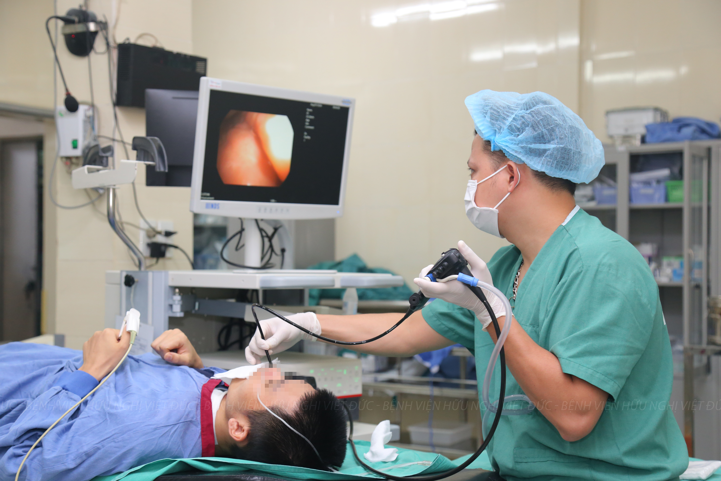
Doctor Nguyen Duc Anh – Department of Neurosurgery II, Viet Duc University Hospital – said: The surgery for the patient lasted nearly 12 hours. Because the tumor is large, doctors had to use modern navigation systems to remove the tumor and with the support of the Harmonic scalpel, reduced the tumor volume and was able to dissect the tumor out of the vascular structure which leaded to the nerves and the base of the skull has been successfully preserved.
The giant meningeal tumor at sphenoidal bone are mostly benign, developing slowly, so the patient can tolerate, until it is detected in a late stage. If it is not controlled and treated in time, the worst scenario is the patient will suffer brain compression, coma, paralysis. When the patient is hospitalized late, the surgery to remove the tumor will take a lot of time and efforts. Meningioma is a common tumor, can be found in many locations: in the anterior, mid or posterior base of the skull. With mesothelioma, depending on the location of the tumor that divide into 3 part including 1/3 outside, 1/3, 1/3 inside.
Doctors recommend that even the tumor is known to be benign, but when the tumor grows, it will compress and cause blurred vision, the patient will have signs such as: transient headache, decreased vision, incorrect words, … it is necessary to find any specialities as soon as possible to be promptly examined, diagnosed, detected and treated in time.


