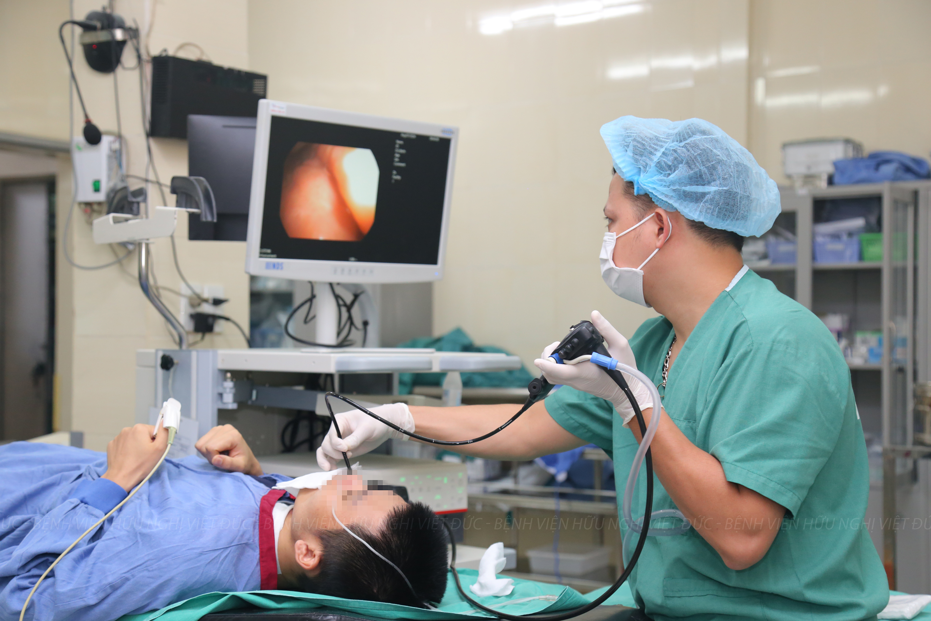
First time in Viet Nam: Arthroscopic reconstruction of wrist joint
28/03/2022 14:05
For the first time in Viet Nam, doctors of Viet Duc University Hospital have successfully performed arthroscopic reconstruction of wrist join for Mr. M.L.A.T, 30 years old, Ha Noi.
Mr. T comes to Viet Duc University Hospital to be examined with pain and instability of his wrist joint, greatly affects to daily living. Mr. T says he had sport injury while playing soccer a year ago, had examination and treatment in many places but couldn’t find the disease. End of January 2022, when playing the skating sport he has injured again, made him very difficult to hold tools, carry, take the motorbike, even picking pen for writing.
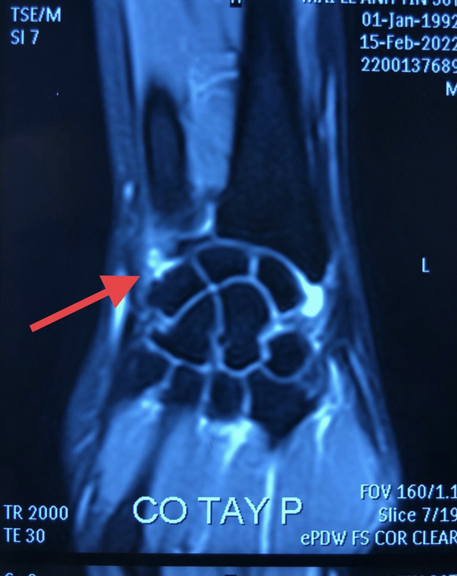
Ass. Prof. Nguyen Manh Khanh – MD, PhD – Vice chairman of Orthopedic and Trauma Institute, Head of Upper Extremities and Sport Medicine Department, Viet Duc University shared: After having examined the clinic manifestations: pain in wrist central area, lost right wrist joint balance, especially the ulnar region, doctors have diagnosed the damage to the triangular fibrocartilage complex. On 2nd March, doctors decided to surgery for patient using arthroscopic reconstruction of wrist joint (Treatment of damage to the cartilaginous triangle ligament complex of the wrist – TFCC-Triangular Fibrocartilage Complex tear). This can be considered as one of the most complicated trauma surgery techniques that performs with laparoscopy, requires in-depth experience, synchronization and modern devices, equipment.
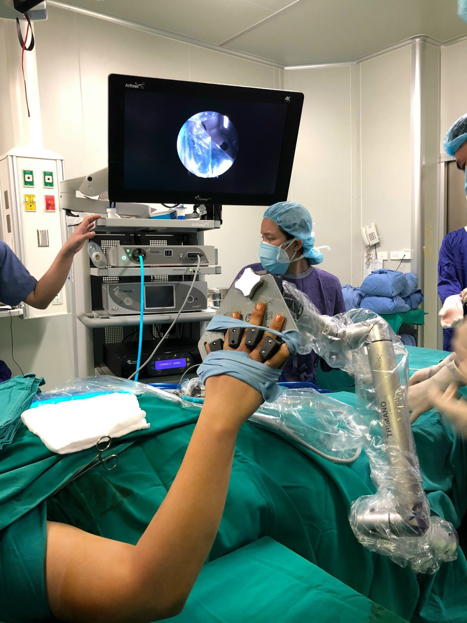
Doctors of Upper Extremities and Sport Medicine Department, Viet Duc University Hospital are operating for Mr. T.
During the operation, surgeons have used specialized arthroscopy set for small joins, in specific is endoscopic tube with 2,7 mm diameter (different with knee, shoulder join which used endoscopic tube with 4,5 mm diameter). Whilst observing found there is inflammation, mild degeneration, damage to the triangular fibrocartilage complex ligament causing instability of wrist join. Surgeons decided to reshape the ligament to help it reconnect, firm and completely by laparoscopy method.
In the past, diagnosis technique was too difficult, some cases were operated on conventionally but with huge surgical incision, destroy soft and bone areas a lots. As the result, using arthroscopy technique has many advantages for patients, small cut (0,5cm), able to fully evaluate within wrist joint about bone, joint, ligament system, helps to treat that lesion properly. Immediately after operation, patient can move slightly wrist joint and less than 24 hours later post-surgery, patient is able to discharge from hospital. After surgery, patient will have to be immobilized with a brace for about a week to stabilize the soft area, reduce wrist edema, then be rehabilitated and after 3-4 weeks, he can return to play sports as normal.
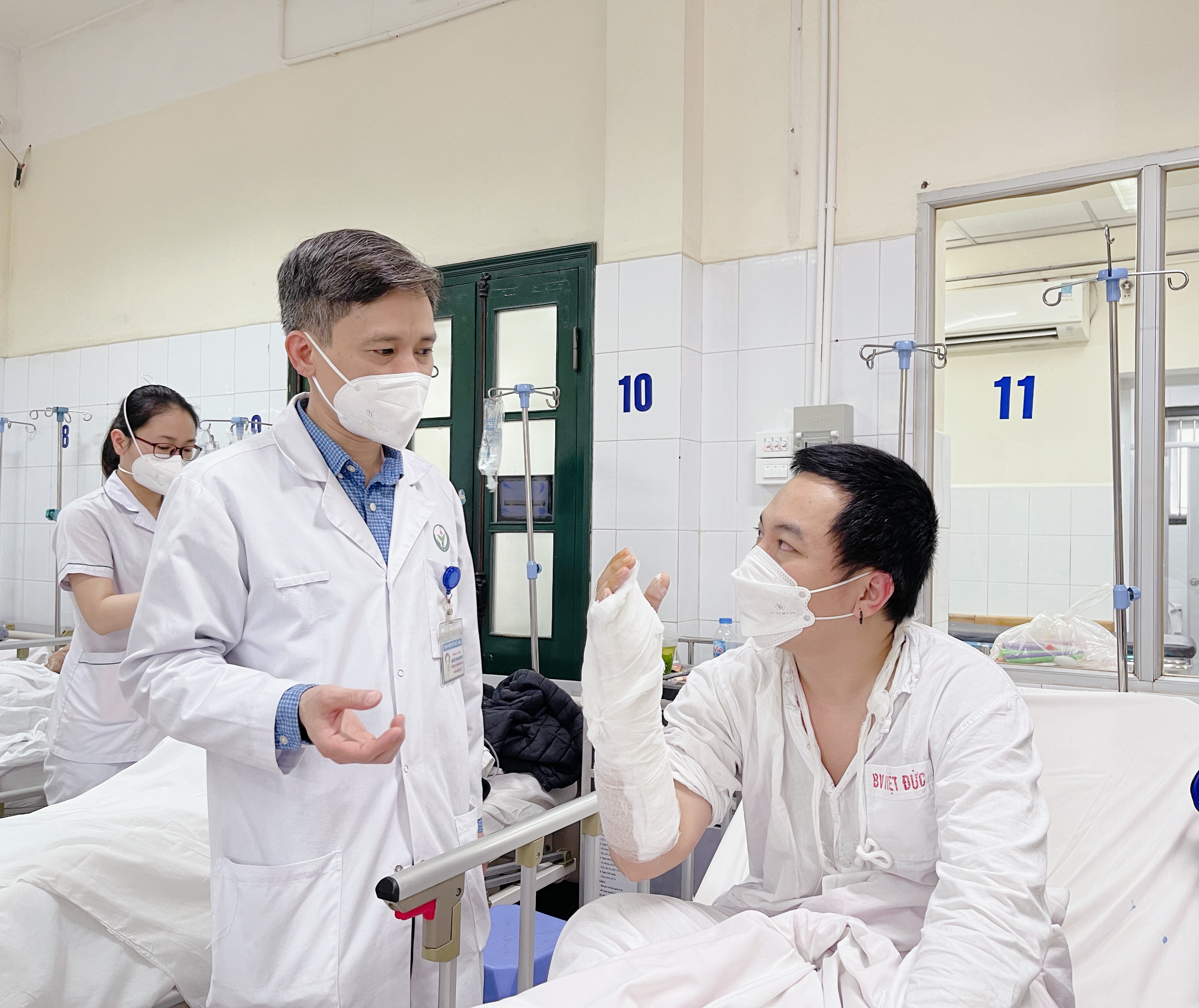
Explaining the reason why this method is only deployed recently in Viet Nam, Ass. Prof. Khanh – MD, PhD says: Knee arthroscopy has become routine and can be performed in many facilities; some facilities also started performing shoulder arthroscopy; Ankle and elbow joints arthroscopy is very rare, mainly in specialized facilities such as Viet Duc University Hospital. Viet Duc University Hospital is the first place in Vietnam to perform arthroscopy wrist reconstruction due to very intensive and complicated wrist joint disease. The wrist is a complex of the lower ends of the two bones of the forearm, with the radial and ulna. Next, there are 8 small bones in the cylinder, which are: Trapezium, trapezoid, scaphoid, lunate, triquetrum, pisiform, capitate, and hamate. Next is a series of metacarpal bones to help movement of the hand more flexible. In order to connect the joints and for the joints to function properly, there are many different, very complex ligament systems.
The diagnosis of pathology is not easy without in-depth knowledge of this specialty, especially in the areas of reconstructive surgery and sports medicine. Injuries like these will be confused with different diseases due to the specific anatomy of many bones, joints, and ligaments, which are prone to injury, especially in sports injuries, greatly affecting living functions, move of patient…so to diagnose of the disease requires a very careful examination. At the same time, along with the clinical diagnosis, imaging is also very important, with ordinary X-ray films it is difficult to detect the disease, it is necessary to rely on specialized imaging techniques, such as CT scan or MRI. In particular, MRI has high value in the assessment, detection and orientation of injuries, especially wrist ligament injuries. Radiologists must also be well trained to be able to detect lesions.
Wrist join is too small, which requires delicate movements as well as specialized, in-depth devices like endoscopic tube, camera, support tools.
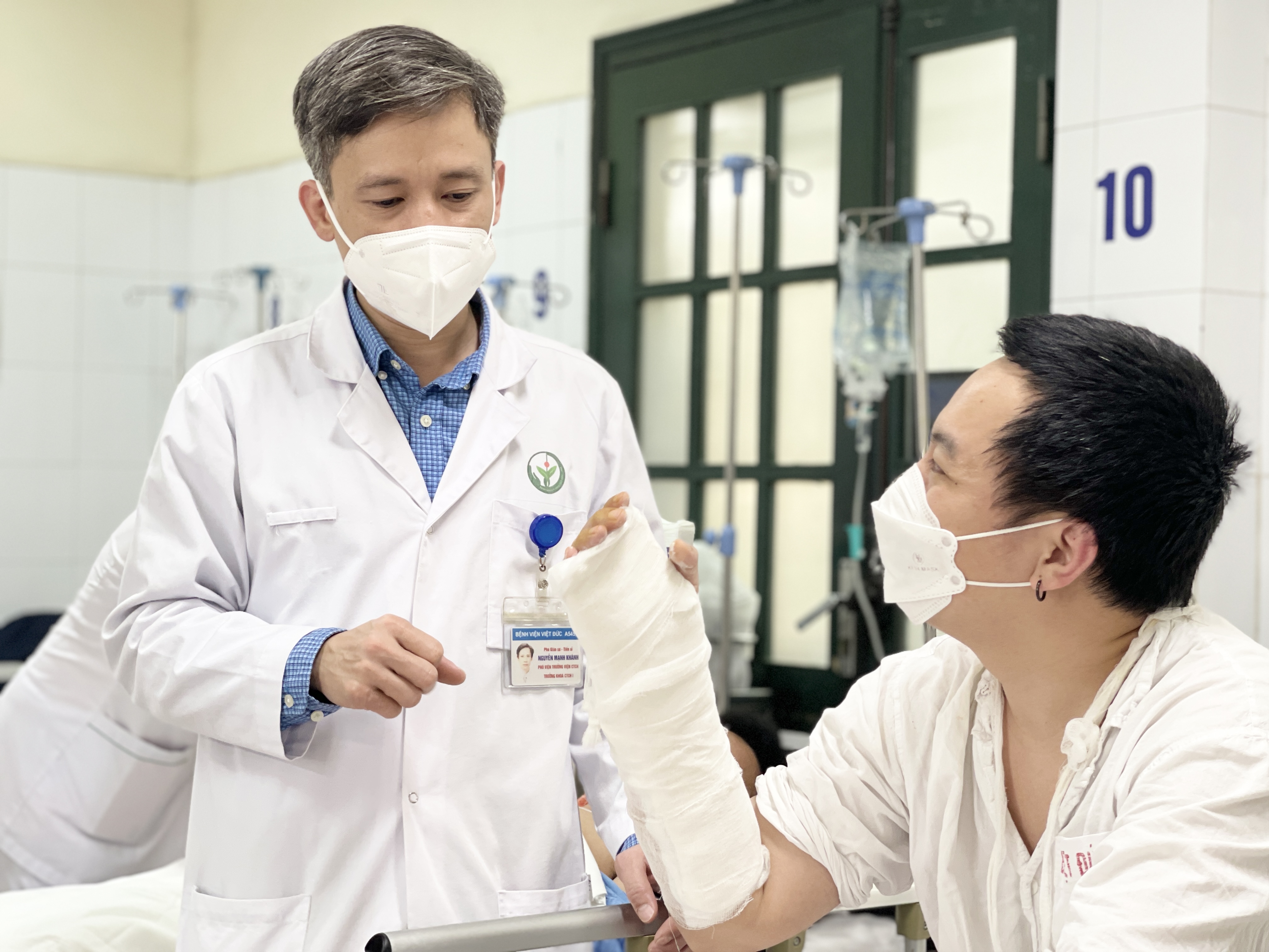
Immediately after operation, patient can move slightly wrist joint and not 24 hours later post-surgery, patient is able to discharge and after 3-4 weeks, patient can return to play sports as normal.
At Viet Duc University Hospital, trauma patients due to sport play are accounted for many, but majority were examined in late, mostly do not think of damages or overlooked due to misdiagnosis, no orientation from the beginning. Hence, doctors recommend: Wrist – hand are the flexible joint of humans, greatly affecting mobility functions not only in sports but also in labor and daily activities. Injury to the wrist joint is one of the most common injuries that is easily overlooked. When suffering an injury, patient should not be subjective but should be examined by specialists to avoid missing the injury and get timely treatment.


