Multiple specialties combination saved a patient with foreign object that deeply plugged the left eye
30/09/2022 10:49
On 17th September 2022, Viet Duc University Hospital took in a case of patient T.P.N (17 years old comes from Phu Tho) admitted to hospital with diagnosis of brain wound, intracranial foreign object, left eyeball wound due to traffic accident motorbike – car. Patient was in emergency at Vietnam National Eye Hospital then transferred to Viet Duc University Hospital in a drowsy condition with lots of scratches, especially a foreign object bar plugged deeply the left eye caused vision loss.
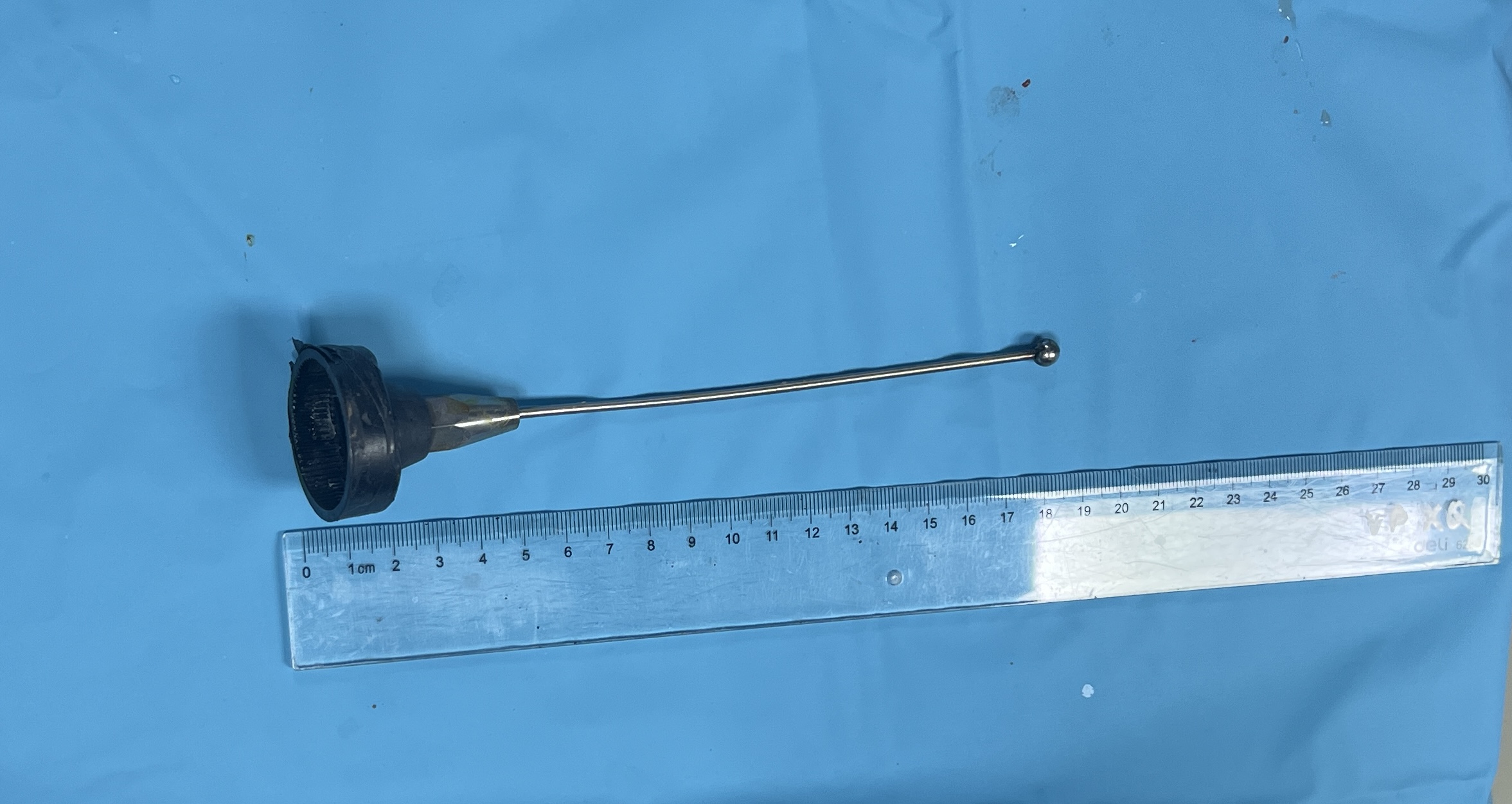
The 2nd degree specialist, Dr. Tran Son Tung, surgeon of Neurosurgery II – neurologist on duty of emergency identified this as a complex case, deep injury and higher risk of vein, neurological damages. He was indicated for multi-slices CT scan in-time emergency to evaluate damages. On the CT scan findings, it showed a foreign object bar of 170x6mm in length, in which the part inside skull was 120mm in length passed from close range of the inner orbit wall through just below the superior wall of the posterior ethmoid sinus on the left of the sphenoid sinus, going close to the medial border of the right internal carotid artery, the cavernous sinus, passes through the pituitary into the brain parenchyma, and passing below the pedicle of large brain part, right border of pons and ends in right hippocampus, next to posterior cerebral artery. The foreign body is also close to the medial border of the right internal carotid artery in the cavernous sinus segment.

Doctors started to remove the foreign object guided by diagnostic imaging, control cerebral bleeding under the increasing light screen.
As soon as the lesion was accurately identified, under the direction of the hospital leader, multi-specialty consultation was held with the participation of specialties: neurosurgery, anesthesia resuscitation, diagnostic imaging, thoracic cardiology. All options as well as risk of complications are discussed thoroughly among leading experts. The indication given is to remove the foreign body under the guidance of diagnostic imaging, control cerebral bleeding under the increasing light screen.
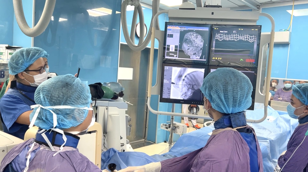
The patient was transferred from the emergency area to the vascular intervention room, taken the endotracheal anesthesia and catheterization from the femoral artery to the cerebral arteries, control of the blood vessels lying in the path of the foreign object. The process of removing foreign object was conducted by the Neurosurgery team led by Assoc. Prof. Ngo Manh Hung, MD, PhD – Deputy Director of Neurosurgery Center, Deputy Head of Neurosurgery Department 2 in charge; together with the coordination of the vascular intervention team led by Dr. Le Thanh Dung, MD, Phd – Head of Diagnostic Imaging Department, under the close supervision of anesthesiology resuscitation team led by Dr. Luu Quang Thuy, MD, PhD – Director of the Center for Anesthesia and Surgical Resuscitation that mainly responsible for.

The foreign object was removed without any vascular damage on the examination film right on intervention table
With the high concentration of all experts, the manipulations are carried out carefully and meticulously to minimize the possible risks, the foreign object was removed without any damage to vessels on the screening film right on intervention table. The patient was then continued to be monitored and resuscitated at the Center for Anesthesia and Surgical Resuscitation, indicated for emergency surgery to suture the sclera wound as soon as the craniocerebral condition stabilized.


